Why people leave their bodies to medical research
UK – Helping find answers to hereditary diseases, training surgeons or wanting to leave an educational legacy are the reasons. But what sort of people donate bodies to science? Neurosurgery at a London hospital.
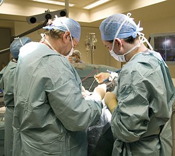
he first thing you notice as you walk into the operating theatre is the smell. There isn’t one. There’s no whiff of chemical preservatives, no sniff of disinfectant and, reassuringly, no smell of bodies. The room is brightly lit, spotlessly clean and there’s little noise apart from the buzz of air-conditioning units, the clatter of surgical tools and hushed conversation.
It takes a while to get your bearings in such as clinical environment. It’s only when you do that you notice the dismembered human arms lying on bloodstained white absorbent pads on the stainless steel operating tables. Some have been severed above the elbow, others at the shoulder. Their waxy flesh is bruised – a natural process caused by the settling of blood after death – and their palms are covered with purple pen marks and deep incisions. Pairs of trainee surgeons in blue gowns and latex gloves work on each arm, supervised by consultants. This morning they dissected the fingers and palms, prising apart the skin to expose garish yellow layers of fat and chalk-white bones. This afternoon they are practising tendon repair.
I’m here as a guest of Vishy Mahadevan, the ebullient professor of anatomy at the Royal College of Surgeons in London and passionate enthusiast for the use of cadavers in medical training. “When I was a surgical trainee more than 34 years ago, it was an apprentice model,” he says over a coffee afterwards. “The only way you could learn was by seeing your boss doing the operation. You assisted them repeatedly and then if your boss was happy you had the basic skills, they would assist you.”
Since the 2004 Human Tissue Act, however, trainee surgeons have been allowed to learn on human cadavers and the college uses 100 bodies each year. Unlike corpses used in undergraduate classes, these are not embalmed but frozen below -20C. Traditional embalming techniques harden tissue and change its texture – fine for exploring anatomy, but hopeless for practising incisions and sutures. So for the surgical training, the bodies are frozen and then thawed before use. Untainted by formalin, the skin and muscle yield to the scalpel like living flesh. The professionalism and respect shown to the donor is impressive. Each arm is tagged with a green label to ensure it is returned to the correct body. At the end of their use, the bodies will be buried or cremated, with ashes returned to their families if they wish. “They are the ultimate simulation model for living humans,” says Mahadevan. “We are so grateful as a profession for the wonderful generosity of individuals who donate their bodies for no other reason than to benefit mankind.”
Tens of thousands have made the decision to leave their body to science, and about 600 do so each year. Even more want to leave their brains for research. Susan Harbot decided to leave her brain to the Parkinson’s UK brain bank six years ago. She had been diagnosed with the disease three years earlier. After her death her brain will be used for hundreds of experiments into the distressing degenerative brain condition. “We have five generations of Parkinson’s sufferers in the family and out of the seven children, three of us have Parkinson’s,” says Harbot, 65, from Horwich, Bolton. “I read about it in a magazine and I didn’t hesitate. I’m not a religious person. At the end of the day it’s my decision and if it helps one person I’d be happy.”
Ed Sykes, 34, a science communicator living in east London, also plans to leave his brain or body to science, and is currently finding out which would be of greater benefit. He says: “I’m not precious about my body. Once I’m dead, I’m dead. It’s either going to feed the worms or be cremated otherwise, so it makes sense to get some use out of it.
“I work in neuroscience and mental health and so I’m interested in leaving my brain to science. I know how significant brain research is going to be in the future. Neurodegenerative diseases are going to be the next cancer – most of us will end up having them. But I also know there’s a shortage of bodies for medical students and until we can recreate an entire body to dissect there’s no substitute for learning from the real thing. If it was possible I’d like to do both – leave my brain to brain research and the rest of my body to medicine.”
Donation is regulated in England and Wales by the 2004 Human Tissue Act, the political response to the Alder Hey organ scandal of the late 1990s when it emerged that hospitals were storing organs from children without consent. It established the Human Tissue Authority to regulate and license removal, storage and use of body parts and organs. Scotland has separate, but similar legislation.Chris Birkett, of the HTA, says: “It boils down to consent. There was a human tissue act before 2004 but it was felt that the act didn’t provide enough safeguards to the public about how tissue was taken and stored for future use.”
Before the act, consent to leave a body to science could be given orally, leaving the potential for abuse or confusion. Now potential donors must sign a witnessed consent form stating what their body will be used for and how long it can be kept: donors can say whether their body can be kept indefinitely or for up to three years. The act allows body parts to be used in medical dissection, surgical training, research and public display. Consent is always needed for research on human tissue from dead people unless the samples were obtained before 1 September 2006. Unclaimed bodies are no longer used. Corpses in the UK are not allowed to be used for road safety testing as they are in the US, and there are no body farms, where bodies are left out to decompose to shed light on forensic science, as there are in the US.
Regulation is onerous for colleges. Every body in dissection in medical colleges must be stored and used in a way that it can be identified. ID tags are attached to every body – on their ears, thumbs and feet. Any body parts removed must be returned to the body, or to a plastic box kept with the body. Alder Hey hangs heavy over medical schools and it crops up in almost every conversation I have with those charged with looking after tissue. According to the HTA, there is no regulatory reason why observers cannot attend, yet none of the medical schools I approached was prepared to open its doors to an undergraduate dissection.
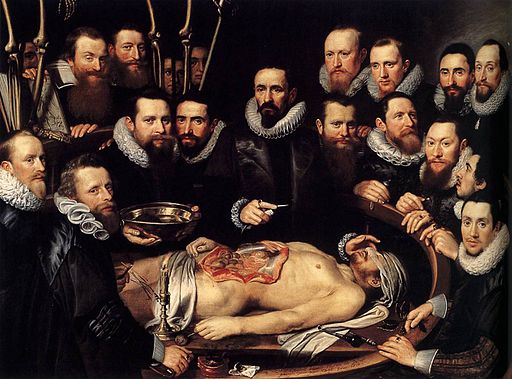
This squeamishness stands in contrast to the early days of modern medical dissection. When Andreas Vesalius published De Humani Corporis Fabrica, his seven books on the structure of the human body, in 1543, crowds paid to see the corpses of criminals cut up in public. The educational benefit of these displays was limited and was often as much about punishment as medicine. In England, dissection was linked in the public minds with dishonour and desecration of the soul so much so that high-level crimes became “punishable by dissection” in the 1752 Murder Act. As anatomy classes flourished, so did the need for bodies, fuelling the rise of the 18th- and 19th-century grave robber. The infamous duo of Burke and Hare went further, murdering multiple victims to cash in on the demand. In response to pressure from doctors, the UK passed the 1832 Anatomy Act. For the first time anatomists needed Home Office licences and were inspected. Unclaimed corpses could be used – and for the first time people could legally donate their body to medical science.
At Cambridge today, the medical school uses 48 cadavers a year. Donors are usually in their 80s or 90s when they die. “There is no typical donor,” says Dr Michelle Spear, deputy clinical anatomist at the university. “One thing they have in common is that they are generous. It’s an altruistic gift and we appreciate it enormously.” The decision to donate may belong to the individual, but it impacts hugely on family. Medical schools and brain banks rely on families to tell them if a donor dies and potential donors are encouraged to discuss their wishes with their next of kin. Brains have to be collected and removed within two days, and only after a death certificate has been produced. They are removed in such a way not to be visible in an open coffin.
Body donations can be even more disruptive. A medical school has to accept a body within six days of death, although typically it is embalmed within three. Without a body, there can be no funeral. It may be two years before the ashes or remains are returned for burial. At Cambridge, bodies are taken by undertakers to the anatomy department where they are washed. Embalmers then pump embalming fluid, a mixture of ethanol and formalin, into the arteries. Typically 25 to 40 litres is used. Bodies are refrigerated and checked regularly to see that the process is working. Additional fluid can be injected directly into areas that are not responding. Once preserved, bodies used for teaching are kept on a metal table in the temperature controlled dissection room and covered with a plastic “dignity shroud”, with another sheet over that. Students work in groups of six or seven on the same body over the their first year, dissecting twice a week in two-hour sessions. Dissections are guided by demonstrators, often retired surgeons.
Students know little about their donor, other than the age, occupation and cause of death. For many students, it’s their first encounter with a corpse. They are eased into their first dissection with videos, photographs and discussions. “In terms of the journey our students take, they have a lot to reconcile from their first dissection onwards,” Spear says. “They might start thinking about mortality – they are confronting death. There are issues of privacy because the subject is naked and so on.”
Spear insists the stories surrounding medical students that deter some people from donating – taking bodies on pub crawls, the hands left in taxis – are myths. “In addition to strict HTA regulations, students sign a code of conduct committing to observe the behaviour appropriate to someone entering the medical profession and we have never had an issue,” she says. “However, I simply do not think that the type of behaviour described by these myths is something that our students would ever want to do. We refer to our donors in a respectful manner and encourage students to think of them as silent teachers.” At the end of the academic year, students attend a committal service where they learn the donor’s name and are shown letters, newspaper clippings, even drawings by their grandchildren. The university also holds a bi-annual memorial service in King’s College chapel and invites family members.
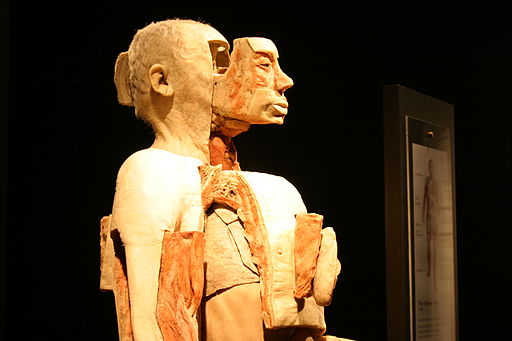
Body Worlds exhibition in San Diego, photo by Patty Mooney (wiki creative commons)
Noel Jackson, who has known since early childhood that he wanted to give his body to science, has no concerns about how his remains will be treated. A former secondary school science teacher and head of education at Newcastle’s Centre for Life, he sees donation as a natural extension of his vocation. Last year, while preparing to host Gunther von Hagens’ Body Worlds Vital exhibition at Newcastle – a display of dissected human corpses preserved using a process called plastination, he decided to let his body be used in a future touring exhibition. “My family has given bodies to science going back at least three generations,” says Jackson, 58. “I’m not a religious man. When you are gone, you are gone. I’ve been a science teacher all my life and I’ll be a science teacher when I’m dead. Some of the people who see Body Worlds will be inspired to take up careers as scientists and doctors. And if I can help encourage that after I’m dead, that’s a worthwhile thing.”
Body Worlds artfully straddles the line between education and entertainment.When it first came to London in 2002, it generated controversy for the way the bodies – skillfully preserved by replacing the water in cells with resin and then artfully dissected – were arranged.
More than 40 million people have seen a Body Worlds show worldwide; 180,000 people saw the most recent in Newcastle. The show features all von Hagens’ trademark qualities. It is thought provoking, technically accomplished and playful. At the entrance, visitors encounter a skeleton in a running pose handing a baton to a figure made of soft tissue. On closer examination, both figures turn out to be from the same donor. Another body was dissected in the pose of a fisherman with hundreds of body parts suspended in mid air on fishing lines, a version of the “exploded” diagrams normally seen in a children’s Dorling and Kindersley science book. It says something about the human response to corpses that the atmosphere in the exhibition was cathedral like. Outside the voices of children filtered through from the nearby cafe. But inside, among the bodies and tasteful dark drapes, tones were muted. At the exit is a consent form, filled in by an anonymous donor – a reminder that these are not plastic mannequins, but once living people. Von Hagens has no shortage of donors. His exhibitions have used 1,100 bodies – but he claims to have another 12,100 living donors signed up. One is Emma Knott, a PR consultant in London. “I was so inspired after I saw the exhibition], which is why I made that decision,” she says. But does she have reservations? “Not really, I mean let’s face it I’m going to be dead.” For her, the attraction lies in encouraging people to get excited about science and anatomy. “The bodies looked so incredible and beautiful and I just thought that would be a fantastic thing to leave once you have left the world – to be preserved in that fashion.”
The HTA says there is no shortage of bodies for more traditional forms of medical science either, although some regions could do with more donors. That’s partly because medical schools are turning their backs on traditional whole-body dissections. Most British universities such as Newcastle teach anatomy using prosections, body parts that have been expertly removed by technicians. Dr Debra Patten, director of anatomy and clinical skills at Newcastle University, says: “One school of thought is that there is a finite amount of time and when other important things have been introduced into the curriculum such as communication skills and medicine in the community, other things have to go out. Having a prosection that’s been expertly prepared is an effective use of time and resources.” Another reason given is that in a group of six or seven students round a cadaver, one only or two will be dissecting at any one time. “With a prosection it’s easier to make sure everyone sees everything,” says Patten. “I did dissection myself as a student, and thoroughly enjoyed it. But I would probably have learned more from prosection.”
Some medical schools have gone further. Plymouth uses no human bodies in its anatomy classes, teaching students instead from live volunteers, mannequins, computer images and data from scans. At Cambridge, Spear is unconvinced that doctors can learn without a body. Dissection builds spatial skills, manual dexterity and sense of touch. It also promotes teamwork, professionalism, communication and helps students cope with death. “There are some things that textbooks cannot provide,” she says. “I wouldn’t take my car to a mechanic who had only read a Haynes manual and worked on a simulated engine.’’ Dissection also toughens them up. Angharad Everden, 23, in her fifth year of medical training at Cambridge University, describes the donation of a body to science as a gift – and says that her first tentative steps into the dissection room were daunting. “These sessions were demanding and made me reflect on death and dying in a I way I hadn’t previously,” she says. “These feelings were quickly replaced with a sense of responsibility to learn the intricacies of the human body for myself, coupled with a growing deep respect for the person who had bequeathed this gift.” Only through dissection can students appreciate the overlap between the different systems that make up a body and see the effects of disease first hand, she says. “I won’t forget the surprise at our discoveries of variations in our individual which in turn will serve to remind me how unique each living person is and not let me fall into the trap of stereotyping people.
“At the end of the year, when we found out the names and personal details of the donors, people were crying. It’s an experience that stays with you and it’s a really valuable one. You grow up when you go into the dissection room.”
Donation consent forms are available from medical schools or the London Anatomy Office. For the full list see the HTA website
Training tomorrow’s doctors: what they learn from dissection
1 Brain
Dissecting the brain allows students to appreciate the location of its component parts. It also yields valuable information about the blood supply and the possible consequences of interruption to this in different areas, such as through a stroke.
2 Face
There are more than 40 muscles for facial expression. Their complex arrangement and function, and the distribution of fine and delicate nerves that supply them, can only be understood by dissection.
3 Neck
This is a formidably complex system of nerves, blood vessels and tubular organs such as the windpipe and gullet. Careful and detailed dissection simplifies a student’s understanding of the anatomical arrangement.
4 Chest cavity and lungs
Besides sheltering the heart and lungs, the chest contains the windpipe and its myriad branches that spread in the lung. It is only by dissecting the lungs and the complicated system of branching airways and blood vessels within that students can understand their amazing gas-exchanging functions. The role of the heart is intimately related to its internal and external design. It was through detailed dissection of the heart that William Harvey, in the 17th century, made his momentous discovery about blood circulation.
5 Hand and foot
The hand and foot are models of mechanical engineering. It is only by patient, meticulous and comprehensive dissection that the complex and intricate arrangement of tendons, muscles, ligaments and small bones within the hand and foot can be properly understood and the functions of the muscles and tendons fully appreciated.
6 Knee joint
The knee is the major weight-bearing joint in the body. A detailed dissection allows appreciation of the spatial relationships of various muscles to the knee joint and yields much information about the mechanical roles of ligaments, cartilages and muscles.
The Royal College of Surgeons
Originally published by The Guardian, author David Derbyshire.
Leave a Comment
You must be logged in to post a comment.